Assessing accommodative function at near is a vital component of understanding the myopia profile of your patient. Myopic progression in children and adults can be influenced by binocular vision function. There is a reported association between higher levels of esophoria and accommodative lag at near in myopic children and young adults as compared to emmetropes.1-5 Myopic children and young adults also show insufficient accommodative responses to lens-induced blur,1, 6-8 greater variability in accommodative response9, reduced accommodative facility4, 8 and enhanced accommodative convergence (elevated AC/A ratios) when compared to age matched emmetropes.10-12 There is speculation in the literature regarding accommodative lag prior to onset of myopia – evidence shows higher accommodative lag in progressing myopes, regardless of their starting point of refraction as an emmetrope or myope,4 however other studies do not match this significance.13 Myopia development and progression is multifactorial, though, so it’s important to assess all contributory factors – the myopia profile is designed to guide you through this clinical decision making and communication process.
A recent study from China has shown that children with a lower amplitude of accommodation showed a better myopia control response to orthokeratology over two years, indicating that improving accommodative function may be part of the mechanism.14 Measuring accommodative amplitude through facility gives you a dynamic picture of how well your patient can cope with their accommodative lag – how much stamina they have to maintain near point focus.
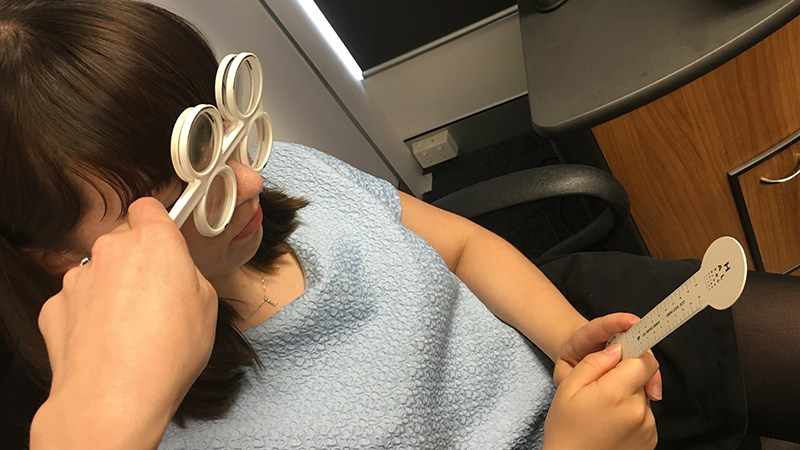
Hand your patient a near point card with N6 or smaller letters – I use the top of an occluder paddle with descending letter sizes, or the back of a Howell phoria card. This should be held at their habitual working distance of 33-40cm. (at the lower end for your shorter armed patients!) Grab your ±1.50 and ±2.00 flippers, and combine the minus sides to give -3.50. If you don’t have flippers yet (and you should order some!) use loose trial lenses held over each eye. You can hold +2.00 and -2.00 in each hand to switch quickly between them for the facility testing described below.
Your paediatric or young adult myopic patient, with their full distance correction in place, should be able to clear this power just as you’d expect their positive relative accommodation / B- test (response to minus) result to reach a similar level. This is a swift measurement of part of the accommodative amplitude response. Then put down the ±1.50 and switch the ±2.00 flippers to the plus side. Again, clearing +2.00 at near is an expected negative relative accommodation / B + test result.15 Flip several times between +2.00 and -2.00 to observe the result, watching for fatigue on one or both sides or if swift clearing is maintained throughout.
Generally agreed clinical protocols for measuring accommodative facility include observation of response times, and if measured by time will include the reaction time of both the clinician and the patient.4 Your result can be qualitative and quantitative. I prefer to record qualitative observations rather than a number of cycles per minute for enhanced clinical detail – for example:
- “Acc fac normal” – if both -3.50, +2.00 and -2.00 are cleared swiftly and consistently
- “Acc fac normal on plus, slow on -3.50, fatigues to fail on -2”
- “Acc fac normal on plus, failed -3.50, cleared -2 on cycles”
If patient 2 or 3 above has a lag of accommodation over +1.00, their results on accommodative facility testing indicate how much ‘petrol in the tank’ they have to account for the lag. Patient 2 is in bigger trouble than patient 3, because they showed an initial response to minus which fatigued to fail, whereas patient 3 consistently maintained lower levels of accommodative function (clearing -2.00). The old adage that there is many ways to skin a cat holds for many clinical techniques as well as record keeping – ultimately we need to know how our patient will functionally cope with accommodative demand through their day and you can adapt facility testing to evaluate this.
Click on this link to read how to measure accommodative lag at near, and this link for advice on prescribing for accommodative dysfunctions.
To delve more into my simplified two-system approach to BV diagnosis and management, you can watch this video entitled Binocular Vision - easier than you think. This one hour lecture includes cases; details easy use of prism correction for vergence disorders; changes to BV in contact lens wear; and why BV matters - for reading and learning in kids, clinical problem solving, and myopia management. You can also download my lecture notes here.
Want to learn more about binocular vision?
Check out my online course Binocular Vision Fundamentals, which starts with my two-system approach to BV assessment and diagnosis. Stepping through understanding of the accommodation and vergence systems, the course then covers clinical tests, diagnostic criteria, prescribing and management. Once this foundation is set, it moves onto clinical communication and the importance of BV in myopia management. Always with a laser sharp focus on the clinical applications.
Included are video examples of assessment techniques and chairside infographic summary downloads to reference in practice.
You can enroll on the first two modules for free, with the full course priced at US$140 if you decide to continue. Reduced course fees by 30% and 50% are available by application for practitioners residing in lower income countries - check out the course page for more information.

About Kate
Dr Kate Gifford is a clinical optometrist, researcher, peer educator and professional leader from Brisbane, Australia, and a co-founder of Myopia Profile.
References
- Gwiazda J, Bauer J, Thorn F, Held R. A dynamic relationship between myopia and blur-driven accommodation in school-aged children. Vision Res. 1995;35:1299-1304. (link)
- Nakatsuka C, Hasebe S, Nonaka F, Ohtsuki H. Accommodative lag under habitual seeing conditions: comparison between myopic and emmetropic children. Jap J Ophthalmol. 2005;49:189-194. (link)
- Drobe B, de Saint-André R. The pre-myopic syndrome. Ophthal Physiol Opt. 1995;15:375-378. (link)
- Allen PM, O'Leary DJ. Accommodation functions: Co-dependency and relationship to refractive error. Vision Res. 2006;46:491-505. (link)
- Price H, Allen PM, Radhakrishnan H, Calver R, Rae S, Theagarayan B, Sailoganathan A, O'Leary DJ. The Cambridge Anti-Myopia Study: Variables Associated with Myopia Progression. Optom Vis Sci. 2013;90:1274-1283. (link)
- Bullimore MA, Gilmartin B, Royston JM. Steady-state accommodation and ocular biometry in late-onset myopia. Documenta Ophthalmologica. Advances In Ophthalmology. 1992;80:143-155. (link)
- Abbott ML, Schmid KL, Strang NC. Differences in the accommodation stimulus response curves of adult myopes and emmetropes. Ophthal Physiol Opt. 1998;18:13-20. (link)
- Pandian A, Sankaridurg PR, Naduvilath T, O'Leary D, Sweeney DF, Rose K, Mitchell P. Accommodative Facility in Eyes with and without Myopia. Invest Ophthalmol Vis Sci. 2006;47:4725-4731. (link)
- Harb E, Thorn F, Troilo D. Characteristics of accommodative behavior during sustained reading in emmetropes and myopes. Vision Res. 2006;46:2581-2592. (link)
- Gwiazda J, Grice K, Thorn F. Response AC/A ratios are elevated in myopic children. Ophthal Physiol Opt. 1999;19:173-179. (link)
- Gwiazda J, Thorn F, Held R. Accommodation, accommodative convergence, and response AC/A ratios before and at the onset of myopia in children. Optom Vis Sci. 2005;82:273-278. (link)
- Mutti DO, Jones LA, Moeschberger ML, Zadnik K. AC/A Ratio, Age, and Refractive Error in Children. Invest Ophthalmol Vis Sci. 2000;41:2469-2478. (link)
- Rosenfield M, Desai R, Portello JK. Do progressing myopes show reduced accommodative responses? Optom Vis Sci. 2002;79:268-273. (link)
- Zhu M, Feng H, Zhu J, Qu X. The impact of amplitude of accommodation on controlling the development of myopia in orthokeratology]. Chinese J Ophthalmol. 2014;50:14-19. (link)
- Evans BJW. Detecting binocular vision anomalies in primary eyecare practice. Pickwell's Binocular Vision Anomalies (Fifth Edition). Edinburgh: Butterworth-Heinemann; 2007:12-38. (link)


If you are interested in topic: make money online selling on ebay & spend
no money upfront – you should read about Bucksflooder first